Salvador-Membreve DM, Jacinto SD, Rivera WL. 2022 Apr 21;8(5):254. doi: 10.3390/gels8050254. Unable to load your collection due to an error, Unable to load your delegates due to an error. Access advice and support for any research roadblock, Full event breakdown with abstracts, speakers, registration and more, Explore the power of knock-out cell lines for your research, crystal-violet-assay-kit-cell-viability-ab232855.pdf. Publishing research using ab232855? Please let us know so that we can cite the reference in this datasheet. Rossitti HM, Dutta RK, Larsson C, Ghayee HK, Sderkvist P, Gimm O. Int J Mol Sci. Dose-response curve of MCF7 (human breast adenocarcinoma cell line) cells to Doxorubicin for 72 hours determined by the Crystal violet Assay Kit (Cell viability) (ab232855). Crystal Violet assay protocol summary:- remove culture medium- wash cells- add Crystal Violet staining solution- incubate for 20 mins- wash cells and air dry plate- add solubilization solution- analyze using a microplate reader at 570 nm absorbance. Chen L, Chiang YC, Chan LS, Chau WY, Lung ML, Kahn M, Lo KW, Mak NK, Lung HL.  Cathepsin B mediated scramblase activation triggers cytotoxicity and cell cycle arrest by andrographolide to overcome cellular resistance in cisplatin resistant human hepatocellular carcinoma HepG2 cells. 2012 Nov;50(8):863-70. doi: 10.3109/13693786.2012.683540. *. For the single cell study, the experiments are performed to analyze the interaction between the individual cell and the substrate, observing the morphological changes, studying the cellular migration, and measuring the traction forces. In the intact organism, cells adhere to a variety of substrates. 2014 Dec;147:33-40. doi: 10.1016/j.exppara.2014.10.003. This site needs JavaScript to work properly. Recommended articles lists articles that we recommend and is powered by our AI driven recommendation engine.
  Adherent cells in laminin-1 wells should be all spread. In a second assay, an aliquot of cells is added to the well of a microtiter plate coated with a test adhesion molecule and the cells are allowed to attach. 3099067 Easy access to products and services you need from our library via powerful searching tools. Population studies involve the analysis of attachment events for a group of cells. There was a highly significant positive linear relationship between crystal violet stained attached cells and the viable cell count of cells attached to aluminium panels (r = 0.9997; p < 0.001: n = 6). Aspirate, add 200 L 10 mg/mL heat-denatured BSA in divalent cation-free PBS and incubate at room temperature for 30 min. Preparation for microdissection (Polyester), Silica-based Plasmid Miniprep (vacuum manifold), Transformation of Frozen Saccharomyces cerevisiae. Cancers (Basel). The amount of Crystal Violet staining in the assay is directly proportional to the cell biomass that is attached to the plate. The https:// ensures that you are connecting to the If I test all of my conditions at RT under the same conditions, will this be a problem?Thanks. If incorrect, please enter your country/region into the box below, to view site information related to your country/region. official website and that any information you provide is encrypted Drug Metab Rev. Would you like email updates of new search results? Invert plate onto an absorbent diaper pad. An official website of the United States government.
  Sudjana AN, Carson CF, Carson KC, Riley TV, Hammer KA. While the blocking is underway, prepare a suspension of the cells to be examined. For fragment E8 or laminin-1 plateau and half-maximal adhesion are usually achieved at 0.5 and 0.05 M, respectively. Count the cell density on a hemocytometer, resuspend the cells to a concentration of 5 x 10. Pull off remaining wash from each well with pipetter. Register to receive personalised research and resources by email. LOXL3 Silencing Affected Cell Adhesion and Invasion in U87MG Glioma Cells. HHS Vulnerability Disclosure. Your browser does not have JavaScript enabled and some parts of this website will not work without it. Contributed by Gordon W. Laurie, School of Medicine, University of Virginia, United States, Cell Adhesion assay protocol from the Laurie Lab, *SN-peptide should be used right after dissolution in ddH2O. It relies on the detachment of adherent cells from cell culture plates during cell death. Store at -20C. Dilute 7.5% BSA (Sigma A8412) to 1% in ddH2O.
  Crystal Violet Assay Kit ab232855 is used for cytoxicity and cell viability studies with adherent cell cultures. Copyright 2022 Creative Bioarray. During the assay, dead detached cells are washed away. Methods Mol Biol. Fix for 10 min at room temp. Before Repeat rock/resuspension prior to removing cell suspension for each row. Invert plate and shake out BSA blocking solution. A simple method to assess bacterial atta . Medicine, Dentistry, Nursing & Allied Health. Pull off remaining wash from each well with pipetter. By closing this message, you are consenting to our use of cookies.
  Please refer to protocols. The incubation time chosen for attachment assay depends on the cell type, as some cells adhere more quickly than others, but 15-20 min is usually adequate.
  Exp Parasitol. Vitale M, Ligorio C, Smith IP, Richardson SM, Hoyland JA, Bella J. Gels.
  Assays are usually performed in triplicate or quadruplicate. Activation of RAS Signalling is Associated with Altered Cell Adhesion in Phaeochromocytoma. The CBP/-Catenin Antagonist, ICG-001, Inhibits Tumor Metastasis via Blocking of the miR-134/ITGB1 Axis-Mediated Cell Adhesion in Nasopharyngeal Carcinoma. Invert plate gently onto an absorbent diaper pad. in a drawer. Add the diluted adhesion molecule to the wells of the microtiter plate (100 L/well). Add coating solution (100 l/well) with multipipetter to wells of a 96 well tissue culture plate (Costar #3595), cover, and place at 4C overnight.
 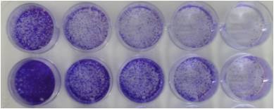 Cell attachment studies include the analysis from the formation of a molecular bond between the cell surface receptors and the complementary ligands (on the surface of ECM) to the observation of a population of cell responses through the cell behavior and changes of morphology during the attachment events. Invert plate gently onto an absorbent diaper pad. Crystal violet is a triarylmethane dye that binds to ribose type molecules such as DNA in nuclei. Pull off remaining wash solution from each well with pipetter. Examine plate in invert microscope.
  my question is this: should i use serum-free media when i seed cells? Paclitaxel, Carboplatin and 1,25-D3 Inhibit Proliferation of Ovarian Cancer Cells In Vitro.
  Invert plate onto an absorbent diaper pad. Cold Spring Harb Protoc. For SN-peptide, use 20 M (MW of SN-peptide is 2412) for plateau or 2.5 - 5 M for half-maximal adhesion. In the first protocol presented in this unit, physical adhesion of a cell is assessed by determining the extent to which the cell spreads on a defined substrate--the plate is coated with the test substance, cells are added and allowed time to attach and spread, and the extent of spreading is assessed using phase contrast microscopy.
  Sci Rep. 2019 Nov 8;9(1):16383. doi: 10.1038/s41598-019-52797-z. 2020 Oct 29;21(21):8072. doi: 10.3390/ijms21218072. Do you normally grow your cells with serum? The site is secure. hi all,i think that perhaps i have been in the lab for too long today and can't get my head to work anymore. 1999-2008 Protocol Online, All rights reserved. To learn about our use of cookies and how you can manage your cookie settings, please see our Cookie Policy. NOT FOR USE IN DIAGNOSTIC PROCEDURES" For licensing inquiries, please contact partnerships@abcam.com, Crystal violet Assay Kit (Cell viability). Invert plate and shake out coating solution. Replace in incubator for 30 min. Pull off remaining coating solution from each well with a yellow tip pipetter.
  Invert plate gently onto an absorbent diaper pad. i guess i'm sort of confused as to how much serum masks the adhesion process (i've been under the assumption that it is needed to assist in adhesion).thanks! Stain for 25 min at room temp. Stain with 100 L 0.1% (w/v) crystal violet, 200 mM MES, pH 6.0 for 60 min at room temperature. Laminin value should be about 1.0 OD. Dilute cells to 2.0 x 10⁵/ml in serum-free medium. Cell proliferation and invasion are regulated differently by EGFR and MRP1 in T-DM1-resistant breast cancer cells. Solubilize dye in 100 L 10% (v/v) acetic acid and incubate on orbital shaker at 150 rpm for 5 min at room temperature. The Crystal Violet assay assumes that all cells that are attached to the plate are "alive" and that all cells that detach are "dead".
 A reliable approach is to weigh out 100 g aliquots with a microbalance for use with individual experiments. For research use only. Clipboard, Search History, and several other advanced features are temporarily unavailable. A common working dilution for the laminin-1 positive control and BSA (Sigma A8412) negative control is 40 g/ml.
 *, *BSA background should be less than 0.1 OD. Incorporation of Natural and Recombinant Collagen Proteins within Fmoc-Based Self-Assembling Peptide Hydrogels. if i use serum-free media, is it also necessary to serum starve the cells? Laurentino TS, Soares RDS, Lerario AM, Marie SKN, Oba-Shinjo SM. Slowly add 100 l/well of serum-free medium down the side of each well. Dose-response curve of HepG2 (human liver hepatocellular carcinoma cell line) cells to Doxorubicin for 72 hours determined by the Crystal violet Assay Kit (Cell viability) (ab232855). Assays were performed according to the kit protocol in triplicate.
  Get resources and offers direct to your inbox. Plateau SN-peptide value is usually 70-80% of laminin. Efficient differentiation of vascular smooth muscle cells from Wharton's Jelly mesenchymal stromal cells using human platelet lysate: A potential cell source for small blood vessel engineering. It depends- are you trying to look at the influence of serum on the cell attachment? *, *Cells in BSA negative control wells should be rare (if not, repeat wash). Read at OD 595. ab232855 has been referenced in 5 publications. Candida albicans adhesion to human epithelial cells and polystyrene and formation of biofilm is reduced by sub-inhibitory Melaleuca alternifolia (tea tree) essential oil. Dose-response curve of MCF7 (Human breast adenocarcinoma cell line) cells to Doxorubicin for 72 hours determined by the Crystal violet staining assay. Examine plate in invert microscope. For the best experience on the Abcam website please upgrade to a modern browser such as Google Chrome. Trichomonas vaginalis induces cytopathic effect on human lung alveolar basal carcinoma epithelial cell line A549. government site. This method is based, with permission, on an original protocol available here. Pull off remaining blocking solution from each well with a yellow tip pipetter. Dissolve/dilute coating substrate in ddH2O at 4C. The adhesion events can be divided into single cell and cell population analysis. Fix cells in the wells to be used for determining 100% attachment value by addition of 100 L 5% (w/v) glutaraldehyde for 20 min at room temperature (or at 4C overnight if necessary). 2016 Apr 1;2016(4):pdb.prot087379. HelloI am doing dayly CV. Please contact us to place your order, or try again later. The cell biomass can be used to infer levels of cell viability / cytotoxicity. Please note: All products are "FOR RESEARCH USE ONLY. 1990;22(2-3):161-78. doi: 10.3109/03602539009041083. With multipipetter, slowly add 100 l/well of freshly diluted 1% glutaraldehyde in PBS. Invert plate gently onto an absorbent diaper pad. Dose-response curve of HepG2 (Human liver hepatocellular carcinoma cell line) cells to Doxorubicin for 72 hours determined by the Crystal violet staining assay.
 Invert plate onto an absorbent diaper pad, then wash plate gently by immersion in a plastic tray containing tap water.
  Assays were performed according to the kit protocol in triplicate. This product is manufactured by BioVision, an Abcam company and was previously called K329 Crystal Violet Cell Cytotoxicity Assay Kit. All rights reserved. With multipipetter, slowly add 100 l/well of serum-free medium down the side of each well (tilt plate; PBS is not recommended for this wash). Not for any other purpose. Photograph selected wells if desired. Activity decreases with storage, even frozen. Figure 1. Review ourcell health assays guideto learn more about our othercell viability,cytotoxicityandcell proliferationassay kits. Federal government websites often end in .gov or .mil. Pour cells in Reagent Reservoir (Costar # 4870), rock to suspend, remove 100 l/well with multipipetter and add to wells. sharing sensitive information, make sure youre on a federal
  Nonattached cells are removed, and the percent of added cells attached to the substrate is quantified by crystal violet staining.
  One more question: how important is the temp and CO2? CABR 2019 Novel Coronavirus Detection FISH Probe, Cell Viability, Proliferation and Cytotoxicity, Fluorescent Cellular Staining Dyes & Ion Probes, Cell-Based Screening and Profiling Services, Pluripotency Verification by Immunostaining, Retinal Pigment Epithelium (RPE) Differentiation, Stem Cell Assay Development and Screening, Epigenetic Induction of Cell Growth Service, Immortalized Cell Line Engineering Service, FISH Probe Design, Synthesis and Testing Service, CAR-T/CAR-NK Target Assessment Service (ISH), CARD-FISH for Environmental Microorganisms (FISH), Transgene Mapping (Locus Amplification & Sequencing), Stable Cell Line Genetic Stability Testing, Animal Chromosome Analysis (G-banded) Service, Digital ISH Image Quantification and Statistical Analysis, Immunohistochemistry (IHC), Immunofluorescence (IF) Service, CYP450 Time Dependent Inhibition (TDI) Assay, Mass Balance, Excretion and Expired Air Collection, Administration Routes and Biofluid Sampling, Animal Models and Testing Services for COVID-19 Research, Humanized-Xenograft Model-based Drug Screening, Syngeneic Tumor Model-based Drug Screening, Cardiovascular Diseases Modeling and Assay Services, Drug-Resistant Cell Model Generation Service.
Adherent cells in laminin-1 wells should be all spread. In a second assay, an aliquot of cells is added to the well of a microtiter plate coated with a test adhesion molecule and the cells are allowed to attach. 3099067 Easy access to products and services you need from our library via powerful searching tools. Population studies involve the analysis of attachment events for a group of cells. There was a highly significant positive linear relationship between crystal violet stained attached cells and the viable cell count of cells attached to aluminium panels (r = 0.9997; p < 0.001: n = 6). Aspirate, add 200 L 10 mg/mL heat-denatured BSA in divalent cation-free PBS and incubate at room temperature for 30 min. Preparation for microdissection (Polyester), Silica-based Plasmid Miniprep (vacuum manifold), Transformation of Frozen Saccharomyces cerevisiae. Cancers (Basel). The amount of Crystal Violet staining in the assay is directly proportional to the cell biomass that is attached to the plate. The https:// ensures that you are connecting to the If I test all of my conditions at RT under the same conditions, will this be a problem?Thanks. If incorrect, please enter your country/region into the box below, to view site information related to your country/region. official website and that any information you provide is encrypted Drug Metab Rev. Would you like email updates of new search results? Invert plate onto an absorbent diaper pad. An official website of the United States government. Sudjana AN, Carson CF, Carson KC, Riley TV, Hammer KA. While the blocking is underway, prepare a suspension of the cells to be examined. For fragment E8 or laminin-1 plateau and half-maximal adhesion are usually achieved at 0.5 and 0.05 M, respectively. Count the cell density on a hemocytometer, resuspend the cells to a concentration of 5 x 10. Pull off remaining wash from each well with pipetter. Register to receive personalised research and resources by email. LOXL3 Silencing Affected Cell Adhesion and Invasion in U87MG Glioma Cells. HHS Vulnerability Disclosure. Your browser does not have JavaScript enabled and some parts of this website will not work without it. Contributed by Gordon W. Laurie, School of Medicine, University of Virginia, United States, Cell Adhesion assay protocol from the Laurie Lab, *SN-peptide should be used right after dissolution in ddH2O. It relies on the detachment of adherent cells from cell culture plates during cell death. Store at -20C. Dilute 7.5% BSA (Sigma A8412) to 1% in ddH2O. Crystal Violet Assay Kit ab232855 is used for cytoxicity and cell viability studies with adherent cell cultures. Copyright 2022 Creative Bioarray. During the assay, dead detached cells are washed away. Methods Mol Biol. Fix for 10 min at room temp. Before Repeat rock/resuspension prior to removing cell suspension for each row. Invert plate and shake out BSA blocking solution. A simple method to assess bacterial atta . Medicine, Dentistry, Nursing & Allied Health. Pull off remaining wash from each well with pipetter. By closing this message, you are consenting to our use of cookies. Please refer to protocols. The incubation time chosen for attachment assay depends on the cell type, as some cells adhere more quickly than others, but 15-20 min is usually adequate. Exp Parasitol. Vitale M, Ligorio C, Smith IP, Richardson SM, Hoyland JA, Bella J. Gels. Assays are usually performed in triplicate or quadruplicate. Activation of RAS Signalling is Associated with Altered Cell Adhesion in Phaeochromocytoma. The CBP/-Catenin Antagonist, ICG-001, Inhibits Tumor Metastasis via Blocking of the miR-134/ITGB1 Axis-Mediated Cell Adhesion in Nasopharyngeal Carcinoma. Invert plate gently onto an absorbent diaper pad. in a drawer. Add the diluted adhesion molecule to the wells of the microtiter plate (100 L/well). Add coating solution (100 l/well) with multipipetter to wells of a 96 well tissue culture plate (Costar #3595), cover, and place at 4C overnight. Cell attachment studies include the analysis from the formation of a molecular bond between the cell surface receptors and the complementary ligands (on the surface of ECM) to the observation of a population of cell responses through the cell behavior and changes of morphology during the attachment events. Invert plate gently onto an absorbent diaper pad. Crystal violet is a triarylmethane dye that binds to ribose type molecules such as DNA in nuclei. Pull off remaining wash solution from each well with pipetter. Examine plate in invert microscope. my question is this: should i use serum-free media when i seed cells? Paclitaxel, Carboplatin and 1,25-D3 Inhibit Proliferation of Ovarian Cancer Cells In Vitro. Invert plate onto an absorbent diaper pad. Cold Spring Harb Protoc. For SN-peptide, use 20 M (MW of SN-peptide is 2412) for plateau or 2.5 - 5 M for half-maximal adhesion. In the first protocol presented in this unit, physical adhesion of a cell is assessed by determining the extent to which the cell spreads on a defined substrate--the plate is coated with the test substance, cells are added and allowed time to attach and spread, and the extent of spreading is assessed using phase contrast microscopy. Sci Rep. 2019 Nov 8;9(1):16383. doi: 10.1038/s41598-019-52797-z. 2020 Oct 29;21(21):8072. doi: 10.3390/ijms21218072. Do you normally grow your cells with serum? The site is secure. hi all,i think that perhaps i have been in the lab for too long today and can't get my head to work anymore. 1999-2008 Protocol Online, All rights reserved. To learn about our use of cookies and how you can manage your cookie settings, please see our Cookie Policy. NOT FOR USE IN DIAGNOSTIC PROCEDURES" For licensing inquiries, please contact partnerships@abcam.com, Crystal violet Assay Kit (Cell viability). Invert plate and shake out coating solution. Replace in incubator for 30 min. Pull off remaining coating solution from each well with a yellow tip pipetter. Invert plate gently onto an absorbent diaper pad. i guess i'm sort of confused as to how much serum masks the adhesion process (i've been under the assumption that it is needed to assist in adhesion).thanks! Stain for 25 min at room temp. Stain with 100 L 0.1% (w/v) crystal violet, 200 mM MES, pH 6.0 for 60 min at room temperature. Laminin value should be about 1.0 OD. Dilute cells to 2.0 x 10⁵/ml in serum-free medium. Cell proliferation and invasion are regulated differently by EGFR and MRP1 in T-DM1-resistant breast cancer cells. Solubilize dye in 100 L 10% (v/v) acetic acid and incubate on orbital shaker at 150 rpm for 5 min at room temperature. The Crystal Violet assay assumes that all cells that are attached to the plate are "alive" and that all cells that detach are "dead". A reliable approach is to weigh out 100 g aliquots with a microbalance for use with individual experiments. For research use only. Clipboard, Search History, and several other advanced features are temporarily unavailable. A common working dilution for the laminin-1 positive control and BSA (Sigma A8412) negative control is 40 g/ml. *, *BSA background should be less than 0.1 OD. Incorporation of Natural and Recombinant Collagen Proteins within Fmoc-Based Self-Assembling Peptide Hydrogels. if i use serum-free media, is it also necessary to serum starve the cells? Laurentino TS, Soares RDS, Lerario AM, Marie SKN, Oba-Shinjo SM. Slowly add 100 l/well of serum-free medium down the side of each well. Dose-response curve of HepG2 (human liver hepatocellular carcinoma cell line) cells to Doxorubicin for 72 hours determined by the Crystal violet Assay Kit (Cell viability) (ab232855). Assays were performed according to the kit protocol in triplicate. Get resources and offers direct to your inbox. Plateau SN-peptide value is usually 70-80% of laminin. Efficient differentiation of vascular smooth muscle cells from Wharton's Jelly mesenchymal stromal cells using human platelet lysate: A potential cell source for small blood vessel engineering. It depends- are you trying to look at the influence of serum on the cell attachment? *, *Cells in BSA negative control wells should be rare (if not, repeat wash). Read at OD 595. ab232855 has been referenced in 5 publications. Candida albicans adhesion to human epithelial cells and polystyrene and formation of biofilm is reduced by sub-inhibitory Melaleuca alternifolia (tea tree) essential oil. Dose-response curve of MCF7 (Human breast adenocarcinoma cell line) cells to Doxorubicin for 72 hours determined by the Crystal violet staining assay. Examine plate in invert microscope. For the best experience on the Abcam website please upgrade to a modern browser such as Google Chrome. Trichomonas vaginalis induces cytopathic effect on human lung alveolar basal carcinoma epithelial cell line A549. government site. This method is based, with permission, on an original protocol available here. Pull off remaining blocking solution from each well with a yellow tip pipetter. Dissolve/dilute coating substrate in ddH2O at 4C. The adhesion events can be divided into single cell and cell population analysis. Fix cells in the wells to be used for determining 100% attachment value by addition of 100 L 5% (w/v) glutaraldehyde for 20 min at room temperature (or at 4C overnight if necessary). 2016 Apr 1;2016(4):pdb.prot087379. HelloI am doing dayly CV. Please contact us to place your order, or try again later. The cell biomass can be used to infer levels of cell viability / cytotoxicity. Please note: All products are "FOR RESEARCH USE ONLY. 1990;22(2-3):161-78. doi: 10.3109/03602539009041083. With multipipetter, slowly add 100 l/well of freshly diluted 1% glutaraldehyde in PBS. Invert plate gently onto an absorbent diaper pad. Dose-response curve of HepG2 (Human liver hepatocellular carcinoma cell line) cells to Doxorubicin for 72 hours determined by the Crystal violet staining assay. Invert plate onto an absorbent diaper pad, then wash plate gently by immersion in a plastic tray containing tap water. Assays were performed according to the kit protocol in triplicate. This product is manufactured by BioVision, an Abcam company and was previously called K329 Crystal Violet Cell Cytotoxicity Assay Kit. All rights reserved. With multipipetter, slowly add 100 l/well of serum-free medium down the side of each well (tilt plate; PBS is not recommended for this wash). Not for any other purpose. Photograph selected wells if desired. Activity decreases with storage, even frozen. Figure 1. Review ourcell health assays guideto learn more about our othercell viability,cytotoxicityandcell proliferationassay kits. Federal government websites often end in .gov or .mil. Pour cells in Reagent Reservoir (Costar # 4870), rock to suspend, remove 100 l/well with multipipetter and add to wells. sharing sensitive information, make sure youre on a federal Nonattached cells are removed, and the percent of added cells attached to the substrate is quantified by crystal violet staining. One more question: how important is the temp and CO2? CABR 2019 Novel Coronavirus Detection FISH Probe, Cell Viability, Proliferation and Cytotoxicity, Fluorescent Cellular Staining Dyes & Ion Probes, Cell-Based Screening and Profiling Services, Pluripotency Verification by Immunostaining, Retinal Pigment Epithelium (RPE) Differentiation, Stem Cell Assay Development and Screening, Epigenetic Induction of Cell Growth Service, Immortalized Cell Line Engineering Service, FISH Probe Design, Synthesis and Testing Service, CAR-T/CAR-NK Target Assessment Service (ISH), CARD-FISH for Environmental Microorganisms (FISH), Transgene Mapping (Locus Amplification & Sequencing), Stable Cell Line Genetic Stability Testing, Animal Chromosome Analysis (G-banded) Service, Digital ISH Image Quantification and Statistical Analysis, Immunohistochemistry (IHC), Immunofluorescence (IF) Service, CYP450 Time Dependent Inhibition (TDI) Assay, Mass Balance, Excretion and Expired Air Collection, Administration Routes and Biofluid Sampling, Animal Models and Testing Services for COVID-19 Research, Humanized-Xenograft Model-based Drug Screening, Syngeneic Tumor Model-based Drug Screening, Cardiovascular Diseases Modeling and Assay Services, Drug-Resistant Cell Model Generation Service.